 rganauta
Eye in 3D
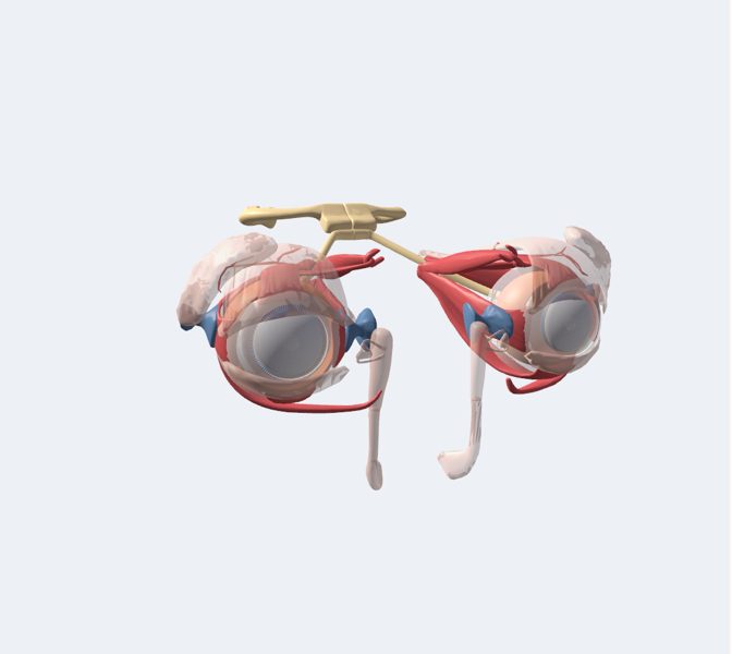
A sphere 24 mm across with three layers: the white sclera with the clear cornea at the front, the uvea (iris, ciliary body and choroid) and the retina.…
Explained for every age
For kids (7–11)
The eye is a living camera. Light comes in through the clear window at the front, the cornea, passes through the little black hole, the pupil, and a lens focuses it onto the screen at the back, the retina, which turns it into signals for the brain.
For teens (12–16)
A sphere 24 mm across with three layers: the white sclera with the clear cornea at the front, the uvea (iris, ciliary body and choroid) and the retina. The cornea and the lens focus an upside-down image onto the retina; cones see color and detail, rods see in dim light. The optic nerve carries the signal to the brain. Six muscles move each eye.
Pre-Med (17+)
Eyeball 24 mm in diameter. Fibrous tunic (sclera, cornea: 43 D of power), vascular tunic (choroid, ciliary body with ciliary muscle for accommodation, iris with pupillary sphincter and dilator) and neural tunic (retina: photoreceptors, bipolar cells, ganglion cells; macula and fovea with cones; optic disc without photoreceptors). Aqueous humor (anterior and posterior chambers, 15 mmHg) and vitreous body. Extraocular muscles innervated by CN III, IV (superior oblique) and VI (lateral rectus). Visual pathway: optic nerve, chiasm, optic tract, lateral geniculate body, optic radiations, occipital cortex.
What you can explore
Tarsi (tarsal plates)Anterior chamber of eyeballAnterior segment of eyeballCorneaIrisLacrimal canaliculusLacrimal glandLacrimal sacLensNasolacrimal ductPosterior segment of eyeballRetinaSclera and corneaIris and ciliary bodyLens and vitreous bodyRetina
Missions
- The living camera Explorer
- Focusing and moving Discoverer
- Visual pathway and clinical practice Pre-Med