 rganauta
Medical imaging in 3D
Diagnostic imaging tests create pictures of the inside of the body to see organs, bones, blood vessels and tissues without surgery. They help find the…
Explained for every age
For kids (7–11)
Medical images are very special pictures of the inside of your body. They let the health team see your bones and organs without having to open anything. Most of them don't hurt: you just have to stay still for a little while.
For teens (12–16)
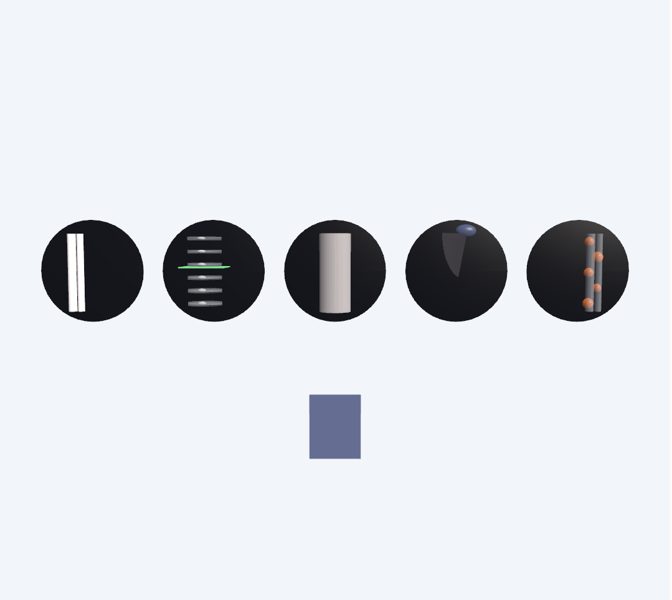
Diagnostic imaging tests create pictures of the inside of the body to see organs, bones, blood vessels and tissues without surgery. They help find the cause of a symptom, catch diseases early, follow how an injury is healing and guide treatments. Each technique uses something different: X-rays, magnets and radio waves, sound, or small amounts of radioactive substances. The team chooses based on the symptom and the part of the body it needs to look at.
Pre-Med (17+)
Diagnostic imaging groups modalities based on different physical principles: X-ray attenuation (radiography and computed tomography), proton resonance in a magnetic field (MRI), ultrasound reflection (sonography) and emission of radiation from radiopharmaceuticals (scintigraphy, SPECT and PET). The first three mainly show anatomy; nuclear medicine shows function (perfusion, metabolism, uptake). Radiography, CT and nuclear medicine use ionizing radiation, so their indication is justified and exposure is optimized; MRI and ultrasound do not use it. The choice depends on the clinical question, the tissue (bone, soft tissue, flow) and the patient (pregnancy, implants, kidney function if contrast is used).
What you can explore
X-ray imaging (radiography)Computed tomography (CT scan)Magnetic resonance imaging (MRI)Ultrasound imaging (sonography)Nuclear medicine (scintigraphy and PET)Radiation safety
Missions
- Looking inside without opening Explorer
- Which exam would you choose? Discoverer
- Physics and clinical judgment Pre-Med